Read the public excerpt
The same instruction and worked examples appear here and in the printable PDF excerpt. You can request full guides for upcoming units below.
READ THIS FIRST
About this guide. Unit 2 carries 10–13% of the multiple-choice weighting and the course’s first real calculations, its first graphs, and its most-run laboratory. The claim it exists to teach is the one Unit 1 set up: a cell is a system of compartments, and the membrane that makes each compartment decides what crosses it. The exam’s own note on this unit is blunt: students “frequently can correctly identify an organelle but fail to accurately describe its function.” Identification is recall; this guide is built for the description.
Aligned to Unit 2 of the 2025–26 Course and Exam Description, Topics 2.1 through 2.10, and to nothing else: every topic is taught here, and the line under each problem’s title names the topics it works. Surface area to volume and water potential live here, with the reference sheet’s formulas and the three it does not give — molarity, dilution, and percent change. Start with the problem that matches your need, open only the relevant Toolbox card, and attempt it before reading its solution. Return to the decision tree or symptom map when you can name the place you are stuck. The rail atop each problem says “Before you compute” — in this unit, that is often literal.
Diagnostic Decision Tree
HOW TO READ THE PROMPT
Unit 2 prompts come in four shapes, and the first job is to name which one you are holding. Use this as a pass through the prompt, not as a stop-at-the-first-match tree.
1. Is there data, a graph, or a table? Read it first: the variables and their units, the trend, any point that breaks it, and whether error bars are drawn. If you are asked to construct a graph, the checklist is fixed: the right type (line for continuous data, bar for categorical), both axes labelled with units, a sensible scale, the points plotted accurately, error bars where the data supply them, a trend line only when one is warranted.
2. What is the verb? Do each job the prompt actually names.
- Identify / state supply the requested information without elaboration.
- Describe the relevant feature, pattern, or trend, with values and units when the data make them relevant.
- Explain / justify / support how or why, with the evidence and reasoning the prompt asks for. For an organelle: the structure, what the structure allows physically, and the function that follows.
- Predict the direction of the outcome, made unambiguous. If justify is also there, that is a second job.
- Calculate the setup visible, the units carried, the answer linked back to the biology.
- Construct / draw the representation with every label it needs.
3. Is it structure-to-function in disguise? An organelle with a folded membrane, a cell that is small, a membrane that is fluid at low temperature, a molecule that cannot cross. Name the structure, say what it does physically (more surface; a hydrophobic interior; a kink that prevents packing), then the function.
4. Is it a direction question? Water, ions, glucose — which way, and by what mechanism. Name the driving difference: concentration for an uncharged solute, electrochemical potential for an ion, or total water potential for water. Then identify the route and energy source. Passive transport moves down the relevant gradient; active transport requires energy, directly from ATP or indirectly from a coupled gradient.
5. Is a formula involved? The reference sheet gives surface area and volume, water potential (, ), and the statistics. It does not give molarity, dilution, or percent change — those three are yours to know. Write the formula, substitute with units, and say what the number means before you move on.
Last check, whatever the verb was: did you complete the requested job? For an organelle, did you describe the function and not only the name? For a direction, did you say which way and why? For a calculation, are the units on the page? For a graph, are both axes labelled?
Where the Points Are Lost
WHAT THE EXAM REWARDS ON THIS UNIT, AND WHERE CREDIT IS LEFT BEHIND
Four facts, each from the people who write and grade the exam.
1. “Identify” is not “describe.” The Course and Exam Description, on this unit: “On the exam, students frequently can correctly identify an organelle but fail to accurately describe its function. Students should be able to explain the relationships between structure and function on both the subcellular and cellular level.” The habit that earns it: every organelle gets a three-part sentence — the structure, what the structure allows, the function that follows. “The mitochondrion makes ATP” is identification. “The inner membrane is folded into cristae, which multiplies the surface holding the ATP-synthesizing machinery, so more ATP can be made per organelle” is the point.
2. Analogies cost points. The same source: “Avoid using catchy analogies (e.g., cell city) and food-based models because on the exam students tend to write about the analogy without demonstrating an understanding of its underlying concept using appropriate terminology.” The habit that earns it: if the word like is in your sentence, replace the comparison with the mechanism. The grader is scoring lysosome, hydrolytic enzymes, vesicle, concentration gradient — not recycling center.
3. Graphs are scored line by line. “Students should be able to label the independent and dependent variables with units, correctly plot data points with appropriate scaling, and correctly represent the data in question … a line graph should be used for continuous data and a bar graph for categorical data. Students often fail to earn points because they draw error bars incorrectly and fail to use them to draw conclusions about the significance of the data.” The habit that earns it: the checklist in the tree above, in order, every time — and when error bars are given, say what they represent before you use them. Say whether they overlap, too, but say it carefully: bars that do not overlap are strong evidence of a real difference, while bars that do overlap settle nothing on their own.
4. A calculation is three points, not one. “Perform the appropriate calculations with correct units while showing their work and linking the results to a biological process.” A correct number with no units, no setup, or no sentence tying it to the cell leaves points on the table. The habit that earns it: formula, substitution with units, answer with units, one sentence of biology — and the self-check is whether another student could follow your setup and say what your number means for the cell. Exam-wide, the last point of a question is the hardest: on one 2025 nine-point question, 2% of students earned the final justify point, so the linking sentence is where a session begins.
Where to Look When You’re Stuck
SYMPTOM FIRST MOVE WORKED MODEL
Symptom | First move | Where |
|---|---|---|
I can name the organelle but not explain it | Structure what the structure allows function, as one sentence. | p. 13, Probs. 1, 8 |
My SA:V answer has no units, or the wrong direction | Write and in one variable; divide; keep . | p. 14, Probs. 2–3 |
I cannot tell what crosses the membrane on its own | Ask two things: is it small, and is it nonpolar? Then find the route. | p. 18, Probs. 4–5 |
Facilitated and active blur together | Down the gradient with no energy is facilitated; up the gradient with ATP is active. | p. 18, Prob. 5 |
The water went the wrong way | Write both potentials with signs; water goes to the lower signed value. | p. 20, Probs. 6–7 |
I forgot , or for a salt | Open container: . Count the ions before you touch . | p. 20, Prob. 6 |
I drew the graph and lost the points anyway | Run the checklist: type, axes with units, scale, points, error bars, trend line. | p. 23, Probs. 3, 7 |
I know endosymbiosis happened but cannot defend it | Evidence, then the claim: double membranes, own DNA, bacterial-style ribosomes, division by fission. | p. 22, Prob. 8 |
I have data but cannot defend the claim | Inventory what the design changed, measured, and failed to isolate. | Prob. 7; Stuck Toolkit |
Master Toolbox — Core Tools
THE UNIT, TOPIC BY TOPIC — WHERE EACH ONE LIVES IN THIS GUIDE
CED topic | Toolbox card | Problems |
|---|---|---|
2.1 Cell structure and function | The parts, one sentence each; Organelles as a system | 1, 8 |
2.2 Cell size | Surface area and volume | 2, 3 |
2.3 Plasma membrane | The membrane, drawn honestly | 4 |
2.4 Membrane permeability | The membrane; What crosses, and how | 4 |
2.5 Membrane transport | What crosses, and how | 5 |
2.6 Facilitated diffusion | What crosses, and how | 4, 5 |
2.7 Tonicity and osmoregulation | Tonicity and water potential | 6, 7 |
2.8 Mechanisms of transport | What crosses, and how | 5 |
2.9 Cell compartmentalization | Compartments, and where they came from | 1, 8 |
2.10 Origins of compartmentalization | Compartments, and where they came from | 8 |
— the graphing skill (4.A) | The graphing kit | 3, 7 |
The exam’s own words for what this unit is for: “explain the relationships between structure and function of organelles and cellular components on the subcellular and cellular levels,” and “select the data necessary to solve a problem and use them to perform the appropriate calculations with correct units.” The cards below are those two sentences with the nouns filled in.
THE PARTS, ONE SCORED SENTENCE EACH: STRUCTURE, THEN THE FUNCTION IT ALLOWS
Part | Structure (what to name) | Function it allows (what to explain) |
|---|---|---|
Ribosome | rRNA and protein; no membrane; in all cells | Synthesizes protein from an mRNA sequence. Present in every known form of life — evidence of common ancestry |
Rough ER | Membrane sheets studded with bound ribosomes | Mechanical support and shape; compartmentalizes; carries out protein synthesis for export and membranes |
Smooth ER | Membrane tubules, no ribosomes | Lipid synthesis; detoxification |
Golgi complex | A series of flattened membrane sacs | Correctly folds and chemically modifies new products (glycosylation happens here); packages proteins for trafficking |
Lysosome | Membrane-enclosed sac of hydrolytic enzymes | Digests material; a role in programmed cell death (apoptosis) |
Vacuole | Membrane-bound sac | Plant: one large vacuole holds water and nutrients and maintains turgor pressure. Animal: smaller, more numerous, store cellular materials |
Mitochondrion | Double membrane; smooth outer, highly folded inner | Compartments for the reactions of aerobic respiration; the folds multiply the surface holding the ATP-making machinery, so ATP is made more efficiently |
Chloroplast | Double membrane; in plants and photosynthetic algae | The site of photosynthesis |
Nuclear envelope | Double membrane around the nucleus; part of the endomembrane system | Separates the genetic material from the cytosol |
The right-hand column is the one the exam scores. Read a row aloud as one sentence — “the inner membrane is folded, which multiplies the surface holding the ATP-making machinery, so more ATP is made per organelle” — and you have the shape of every organelle answer in this unit. The next card puts the parts on their route.
ORGANELLES AS A SYSTEM, NOT A LIST
The exam does not ask what an organelle is. It asks what the organelle’s structure lets it do, and it asks the organelles together: a protein made on a ribosome, folded in the ER, modified in the Golgi, and shipped in a vesicle is one story with five characters. The card above is the parts; this is the route.
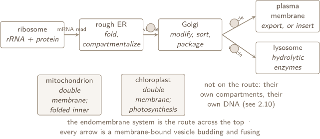
Two sentences to keep. Compartments let incompatible reactions run in one cell: the lysosome’s enzymes work in an acidic interior the cytosol never sees. Folding multiplies surface without adding volume: cristae, and the ER’s sheets, are the same idea as the small cell in the next card.
A check first. One question opens each of the next three cards and is answered where that card ends. Answer it before you read on; getting it wrong is the point, because that is what makes the card stick.
1. SCALE THE MODEL
A spherical cell doubles its radius. By what factor does its surface area change, and by what factor its volume? What happens to surface area per unit volume?
SURFACE AREA AND VOLUME: THE CONSTRAINT EVERY CELL LIVES UNDER
The formulas (given on the reference sheet): cube , ; sphere , ; rectangular solid , ; cylinder , . Divide and the variables mostly cancel: a cube gives and a sphere gives . Notice that the units do not fully cancel — — so the ratio carries units of reciprocal length. It is not a pure number, and it is meaningless unless the area and the volume were measured in the same length unit to begin with.
Why it is a constraint. Everything a cell takes in or gives out crosses the membrane, so supply scales with surface area. Everything a cell consumes or produces happens in the interior, so demand scales with volume. Grow a cell by a factor of two in every direction and its surface grows four times, its volume eight: each unit of interior now has half the membrane serving it. That is why cells are small, why a large cell is folded or flattened or long, and why the CED’s illustrative examples — root hairs, gut epithelial cells, cilia, guard cells, stomata — are all surface-area answers to a volume problem.
The same rule, applied to whole organisms. As an organism gets bigger its surface-area-to-volume ratio falls, so it exchanges heat with its surroundings proportionally more slowly: a small mass loses heat fast, a large mass slowly. The CED’s consequence is in its own words: “typically, the smaller the organism, the higher the metabolic rate per unit body mass” — a mouse burns far more per gram than a horse because it is losing heat far faster per gram. Problem 3 puts numbers on it.
CHECK 1 — SCALE THE MODEL
Area by , volume by , and the ratio by — it halves. You can read that straight off the exponents without computing a single area: area scales with the square of a length and volume with the cube, so their ratio scales with , which is exactly the the sphere formula gives. Doubling halves . That is the whole constraint of this card in one line, and it is why the cell that doubled its radius now has half the membrane per unit of cytoplasm to feed it. Reach for the exponents first; computing and separately gets the same answer and hides the reason.
THE MEMBRANE, DRAWN HONESTLY
Every drawing of a membrane on the exam is scored on the same handful of facts, and they all follow from Unit 1’s phospholipid: a polar, phosphate-bearing head and two nonpolar tails.
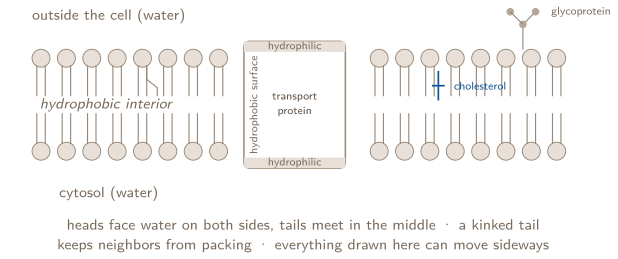
The five facts the drawing carries (2.3.A, 2.3.B):
- Orientation. The polar, hydrophilic phosphate regions face the aqueous exterior and interior; the nonpolar, hydrophobic fatty-acid regions face each other in the membrane’s interior. That is the whole reason it is a bilayer and not a sheet.
- Proteins sit where their chemistry sits. An embedded protein’s hydrophilic regions are either inside the protein or exposed to the cytosol; its hydrophobic regions form the surface that touches the fatty-acid tails. A protein does not float in the membrane by accident — its own polar and nonpolar side chains (Unit 1, Topic 1.7) place it.
- Fluid mosaic. The framework is phospholipid; embedded in it are proteins, steroids (cholesterol, in vertebrate animals), glycoproteins, and glycolipids — and all of them can move around within the membrane. Fluid is the movement; mosaic is the mix.
- Selective permeability is the interior. The hydrophobic core is what stops ions and large polar molecules (2.4.A). The next card is the sorting rule.
- The cell wall is a different structure with a different job (2.4.B): in bacteria, archaea, fungi, and plants it is a boundary outside the membrane that gives structure, is a permeability barrier to some substances, and protects against osmotic lysis — the walled cell in pure water swells and stops; the animal cell bursts.
2. CHECK THE BOUNDARY
A positively charged ion is more concentrated outside a cell than inside. Is that enough to decide whether its entry is passive?
WHAT CROSSES, AND HOW: THE SORTING RULE FOR EVERY TRANSPORT QUESTION
Two questions sort every molecule: how big is it, and is it polar or charged? Then one more decides the energy: which way is it going relative to its gradient?
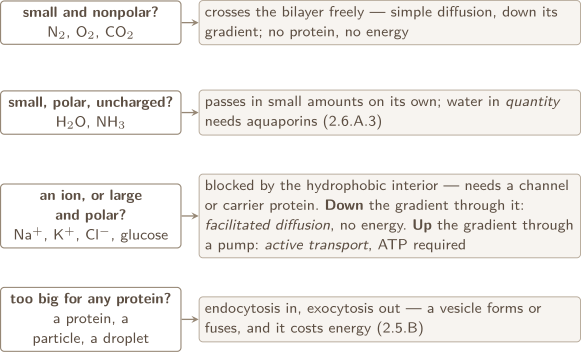
The definitions in the CED’s words (2.5.A): passive transport is the net movement of molecules from regions of high concentration to regions of low concentration without the direct input of metabolic energy; active transport requires the direct input of energy, and in some cases moves molecules from low concentration to high. Facilitated diffusion is passive — the protein provides a route, not a push. The selective permeability of the membrane is what allows gradients to exist in the first place (2.5.A.1); transport is what the cell does with them.
Ions carry charge, so their movement makes a voltage (2.6.A.1, 2.8.A.1). Charged ions such as Na and K need channel proteins to cross, and when they cross, the membrane becomes polarized. The Na/K pump uses ATP to push 3 Na out and pull 2 K in per cycle — against both gradients — and that unequal exchange, plus the gradients it maintains, is what keeps the membrane potential. Membrane proteins are necessary for every active transport event; there is no pump without a protein.
The one-line test, and the exception that breaks it. For an uncharged solute the test is short: find the concentration gradient, then ask only whether it needs a protein — and answer that from size and polarity. For an ion it is not enough. Concentration is one half of an ion’s driving force; the voltage across the membrane is the other, and the two together are the electrochemical gradient. So an ion can move from low concentration to high without any energy being spent, through a channel, when the electrical pull is the stronger half. “Low to high, therefore a pump” is true of glucose and false of potassium, and the exam knows the difference. Active transport is the case where metabolic energy moves cargo against its relevant gradient — the sodium–potassium pump spends ATP directly.
CHECK 2 — CHECK THE BOUNDARY
No, and the reason is the word positively. An uncharged solute is decided by its concentration gradient alone, so “more outside than inside” would settle it: entry is down the gradient, therefore passive. An ion answers to two gradients at once — the concentration difference and the voltage across the membrane — and the prompt gave you only one of them. Most animal cells are negative inside, which would pull a cation in and add to the concentration push; but a cell held positive inside could oppose it hard enough that entry costs energy. Ask for the membrane potential, or say plainly that the answer depends on it. Naming what you were not given is worth more than a confident guess.
3. NAME THE DRIVING DIFFERENCE
Two compartments are separated by a membrane permeable to water. One holds more dissolved solute — but it is also under higher pressure. Is the solute concentration on its own enough to predict which way water moves?
TONICITY AND WATER POTENTIAL: THE DIRECTION, THEN THE NUMBER
Tonicity is a comparison, not a property. An external solution is hypotonic to a cell if it has fewer dissolved particles than the cell’s interior, hypertonic if more, isotonic if the same. Water moves by osmosis from hypotonic toward hypertonic — from the side with more water per particle to the side with less. In the CED’s other phrasing (2.7.B.2): water moves from regions of low osmolarity or solute concentration to regions of high osmolarity or solute concentration.
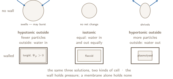
Water potential is the same statement with a number on it (given on the reference sheet): and . Water potential compares water’s tendency to move. Water runs downhill in the way a ball runs downhill in height, and net movement stops when connected regions reach equal potential. Pure water in an open, unpressurized container is defined as zero. Dissolved solute lowers the solute term below zero, while positive pressure can raise total above zero. Negative values are therefore common, not automatic errors.
- — water potential, in bars. Pure water in an open, unpressurized container is .
- — pressure potential. Zero in an open container (the sheet says so in print); positive in a turgid walled cell.
- — solute potential. Always : adding solute only ever lowers water potential.
- — ionization constant: for sucrose and glucose, for NaCl, for . Count the ions.
- — mol/L. Lbar/(molK). — kelvin C .
Units check: . The direction rule: water moves from higher to lower . Compare signed values, never magnitudes; when pressure potential is positive, the lower value need not be negative.
Molarity and dilution (not on the sheet — memorize):
Osmoregulation is the organism keeping this under control (2.7.B): growth and homeostasis depend on the constant movement of water across membranes, and osmoregulation keeps water balance and internal solute composition where the organism needs them. The CED’s two illustrations are worth having ready: the contractile vacuole of a freshwater protist, pumping out the water that constantly enters a hypotonic pond; and the central vacuole of a plant cell, whose stored water and solutes hold the pressure potential that keeps the plant upright.
CHECK 3 — NAME THE DRIVING DIFFERENCE
No. Water moves from higher to lower , and — two terms, and the prompt has given you one of each pulling opposite ways. More solute lowers and pulls water in; higher pressure raises and pushes water out. Which wins is arithmetic, not intuition: add the two signed numbers on each side and compare the totals. The familiar shortcut — water moves toward the side with more dissolved particles — is the special case where the pressure potentials are equal, which is true of two open beakers and false of a turgid plant cell. That is exactly why a walled cell stops taking on water long before its solute concentration matches the outside: has climbed until the sum is level. Say which term you compared, and the direction follows.
COMPARTMENTS, AND WHERE THEY CAME FROM
Why internal membranes (2.9): membranes and membrane-bound organelles compartmentalize the cell’s metabolic processes and specific enzymatic reactions. Two gains, both scored: internal membranes let processes run without competing — an acidic lysosome next to a neutral cytosol, an oxidizing reaction next to a reducing one — and they increase the surface area on which reactions can occur, which is the cristae argument again. A prokaryote typically lacks membrane-bound organelles but is not disorganized: it has internal regions with specialized structures and functions (2.10.A.2). A eukaryote keeps internal membranes that partition the cell into specialized regions (2.10.A.3).
Where two of them came from (2.10.A.1): membrane-bound organelles such as mitochondria and chloroplasts evolved from once free-living prokaryotic cells by endosymbiosis — an ancestral cell engulfed a prokaryote and kept it. When a prompt asks you to support that claim with evidence (skill 6.B), the evidence is structural and it is the same list for both organelles:
- Two membranes — the inner one the prokaryote’s own, the outer one the host’s, from the engulfing.
- Their own DNA, circular, like a bacterial chromosome and unlike the nucleus’s linear chromosomes.
- Their own ribosomes, closer in size and structure to bacterial ribosomes than to the cell’s cytosolic ones.
- They divide by fission, on their own schedule, the way bacteria do — a cell cannot make a new mitochondrion from scratch.
Write the evidence, then the claim it supports; a claim with no evidence attached is a statement, and statements do not score on a 6.B prompt.
THE GRAPHING KIT — SIX CHECKS FOR A CONSTRUCTED GRAPH
The CED lists them (skill 4.A). Check the representation against all six; which of them a particular question pays for is decided by its own rubric:
- 1.
- Type of graph appropriate for the data. Continuous independent variable (concentration, time, mass) a line or scatter graph. Categorical independent variable (species, treatment group) a bar graph. Data spanning several orders of magnitude a log scale on that axis, and say so on the axis.
- 2.
- Axis labels with units, the independent variable on , the dependent on , and a legend if there is more than one series.
- 3.
- Scaling — even intervals that use the space; the origin need not be zero, but the scale must be honest.
- 4.
- Accurately plotted points, each one where the table puts it.
- 5.
- Error bars when the data supply them. The exam’s own complaint: students “draw error bars incorrectly and fail to use them to draw conclusions about the significance of the data.” A bar of SEM approximates a 95% confidence interval; when two groups’ bars do not overlap, the difference is likely real; when they overlap substantially, you cannot claim it is.
- 6.
- A trend line only when the data warrant one.
Percent change is not on the reference sheet, and the exam asks for it (skill 5.A): . Keep the sign; it is the direction.
PROBLEM 1
Two cells, one system: reading organelles as a division of labor
Topics 2.1 · 2.9
A pancreatic cell secretes large amounts of digestive enzymes into a duct. A skeletal-muscle cell contracts for hours at a time and secretes almost nothing. (a) Predict which of the two cells has more rough endoplasmic reticulum and more Golgi, and which has more mitochondria. Justify each prediction from the structure and function of the organelle. (b) Describe the route a digestive enzyme takes from the instructions that encode it to the duct, naming every membrane-bound structure it passes through and what happens to it at each. (c) A toxin blocks vesicles from budding off the Golgi. Predict two consequences for the pancreatic cell and explain each. (d) A different cell cannot make one of the hydrolytic enzymes that belongs in its lysosomes. Predict what happens inside that cell over time, and explain why compartmentalization is the reason the cell has lysosomes at all.
BEFORE YOU COMPUTE
Every part is one sentence pattern applied to a different organelle: the structure what the structure allows the function that follows. Write it three times in (a) and you have written the answer the grader is looking for. The exam’s own warning about this unit is that students identify the organelle and stop. Do not stop.
WORKING
(a) Match the organelle’s structure to the cell’s job.
More rough ER and Golgi: the pancreatic cell. Rough ER is membrane studded with bound ribosomes; the ribosomes synthesize protein directly into the ER’s compartment, so a cell that exports protein in quantity needs a great deal of that membrane. The Golgi is a stack of flattened membrane sacs where newly made products are folded correctly, chemically modified, and packaged into vesicles for trafficking — the step between “made” and “shipped.” A cell whose job is to ship enzymes by the ton has more of both; a muscle cell, which keeps its proteins, needs far less.
More mitochondria: the muscle cell. A mitochondrion’s double membrane makes compartments for the reactions of aerobic respiration, and its highly folded inner membrane multiplies the surface holding the ATP-synthesizing machinery, so ATP is made more efficiently per organelle. Contraction is paid for in ATP, hour after hour; the cell that spends the most ATP has the most of the organelle that makes it.
(b) The route, one membrane at a time. The enzyme’s gene is in the nucleus, inside the nuclear envelope — a double membrane that separates the DNA from the cytosol. The mRNA leaves the nucleus and is read by a ribosome bound to the rough ER; the growing polypeptide enters the ER compartment, where it is folded. A transport vesicle buds from the ER and fuses with the Golgi complex, where the protein is correctly folded and chemically modified — sugar groups are added here, which is what glycosylation means — and sorted for export. A second vesicle buds from the Golgi, moves to the plasma membrane, and fuses with it; the enzyme is released into the duct by exocytosis. Every hand-off in that sentence is a vesicle budding from one membrane and fusing with another — that is what “endomembrane system” means.
(c) Block the Golgi’s exit and two things follow. First, the enzymes cannot leave: they are made, folded, and modified, but the vesicle that carries them to the plasma membrane never forms, so secretion into the duct falls and finished product accumulates in the Golgi. Second — the consequence students miss — the plasma membrane stops being renewed. Exocytosis does not only deliver cargo; the vesicle’s own membrane joins the plasma membrane when it fuses. A cell that keeps taking membrane in by endocytosis and never sends any back out loses surface area over time. Both consequences come from the same fact: the Golgi is the sorting station, and everything downstream of it depends on vesicles leaving it.
(d) Prediction: the substance that enzyme digests accumulates inside the lysosomes, and the cell fills with it. A lysosome is a membrane-enclosed sac of hydrolytic enzymes; material brought to it by vesicles is broken down there. Remove one enzyme and its substrate is delivered but never broken down, so the lysosomes swell with undigested material and the cell’s function degrades. (This is the mechanism of a family of real inherited disorders — lysosomal storage diseases — which is why a single missing enzyme can be fatal.)
Why lysosomes exist at all: hydrolytic enzymes that work best in an acidic interior would damage the cell if they ran loose in the neutral cytosol. Enclosing them in their own membrane lets the cell run a digestive reaction and keep the rest of its chemistry safe from it — compartmentalization minimizing competing interactions, which is Topic 2.9 in one organelle.
ANSWER
(a) Pancreatic cell: more rough ER and Golgi (protein synthesis, folding, modification, and packaging for export); muscle cell: more mitochondria (folded inner membrane more ATP-making surface the ATP contraction spends). (b) Nucleus ribosome on rough ER (synthesis, folding) vesicle Golgi (modification, sorting) vesicle plasma membrane (exocytosis). (c) Secretion falls and product backs up in the Golgi; the plasma membrane is no longer renewed. (d) The undigested substrate accumulates in the lysosomes; lysosomes exist so acidic hydrolysis can run without damaging the cytosol.
WATCH OUT
The scored error on this unit is the missing middle clause. “The pancreatic cell has more Golgi because it secretes enzymes” names two true facts and no mechanism; “because the Golgi is where products are modified and packaged into vesicles for export” is the point. And never reach for the analogy — the CED says in print that students who write “the Golgi is the post office” tend to demonstrate the analogy rather than the concept.
CONNECTION
The muscle cell’s mitochondria are Unit 3’s whole subject, and the cristae argument — fold a membrane to gain surface without gaining volume — is Problem 2’s geometry applied inside an organelle. The enzymes this cell exports are Unit 1’s proteins, glycosylated in the Golgi: the sugar is why they are glycoproteins.
ABOUT THIS EXCERPT
This is the opening of a 40-page guide: the diagnostic tree, the full Master Toolbox, and the first worked problem. 7 more problems follow in the complete guide, each worked the same way — what to notice before you start, every step shown, and the mistake that problem invites. The complete guide is shared with families during the fit conversation.
Engineering Confidence — engineeringconfidence.one
Source and review notes
What this resource is
Complete browser edition of the public excerpt from an original Engineering Confidence AP Biology unit-reference packet. Every problem and scenario was written for this guide.
- Published
- Last reviewed
- Printable edition
- 16 pages
Course reference: official College Board course page.
Read this public excerpt above, or save the PDF for printing and offline reading.
Download the 16-page PDF excerpt